Online Newsletter Committed to Excellence in the Fields of Mental Health, Addiction, Counseling, Social Work, and Nursing
Showing posts with label memory. Show all posts
Showing posts with label memory. Show all posts
July 01, 2015
Long-term memory formation
What do you think of this article?
NYU scientists find that growth factors that build brains also build memories
New York University
"A team of New York University neuroscientists has determined how a pair of growth factor molecules contributes to long-term memory formation, a finding that appears in the journal Neuron.
"These results give us a better understanding of memory's architecture and, specifically, how molecules act as a network in creating long-term memories," explains the paper's senior author, Thomas Carew, a professor in NYU's Center for Neural Science and dean of NYU's Faculty of Arts and Science. "More importantly, this marks another step toward elucidating the intricacies of memory function, which is vital in the development of cognitive therapies to address related afflictions."
The importance of growth factor molecules (GFs) has long been known. They are critical in building brains beginning in utero and until adulthood. Moreover, over time, it's been established that GFs are "recycled" from brain builders to engineers of long-term memories.
Less clear, however, is how the wide range of GF families, as well as different members within each family, act to help us create these memories.
In working to address this question, the NYU research team, which also included graduate student Ashley Kopec, the study's lead author, and research scientist Gary Philips, focused on two GF families: TrkB and TGFβr-II, which represent two distinct classes of GFs that utilize different types of receptors to exert their actions in the brain.
In their study, the researchers examined GFs in Aplysia californica, the California sea slug. Aplysia is a model organism that is quite powerful for this type of research because its neurons are 10 to 50 times larger than those of higher organisms, such as vertebrates, and it possesses a relatively small network of neurons--characteristics that readily allow for the examination of molecular signaling during memory formation.
Specifically, to produce a form of "threat memory" called sensitization in a simple reflex system of Aplysia, the researchers presented the sea slugs with a pair of mild tail shocks delivered 45 minutes apart--the first to instill a "molecular context" in the neurons of the reflex and the second to use that context to drive molecular mechanisms that are required to form a long-term memory -- and then examined GF activity at both periods, Time 1 and Time 2.
Their results showed differences in the role of these two GF families across two dimensions: time and space.
At Time 1, when the context for the memory is first created, TrkB plays a critical role while TGFβr-II is irrelevant. However, at Time 2, when a long-term memory is actually formed, the roles are reversed: TGFβr-II is active, but TrkB is insignificant.
In addition, the results showed spatial differences.
In Aplysia, the simple neural circuit that mediates the reflex modified by learning is made up of unique sensory neurons and motor neurons. The sensory neurons' cell bodies live in one compartment of the brain while their companion synapses, which pass along signals to other cells, reside in another. In the Neuron study, the researchers found that the TrkB effects are exerted only at synapses while TGFβr-II functions only at the cell body.
Overall the study provides new insights into how different GF families play unique roles both in time and in space, thus helping to elucidate the "when," "where," and "how" of memory formation."
###
The research was supported by grants from the National Institute of Mental Health (RO1 MH 041083, F31 MH 100889).
For more information on memory, please visit Aging and Long Term Care CE Course
March 24, 2014
Brain Region Singled Out for Social Memory, Possible Therapeutic Target for Select Brain Disorders
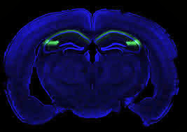
Researchers have found in mice that a formerly obscure region of the hippocampus called CA2 is important for social memory, the ability of an animal to recognize another of the same species. Identifying the role of this region could be useful in understanding and treating disorders characterized by altered social behaviors such as schizophrenia, bipolar disorder, and autism. Funded in part by the National Institute of Mental Health (NIMH), the study was published last month online in Nature.
Background
The hippocampus is essential for learning and memory—specifically the storage of knowledge of who, what, where, and when. Clues about the hippocampus’s roles emerged from the famous case of patient HM (Henry Molaison), who had most of his hippocampus removed by surgeons in 1953 to cure his epilepsy. HM became unable to form new memories of people he subsequently worked with for years.
Most previous studies of how memory is harnessed have focused on the trisynaptic pathway. In this neural circuit, information that is obtained from the entorhinal cortex—the main interface between the hippocampus and the neocortex or the outermostpart of the brain involved in higher functions such as thought or action—proceeds to the dentate gyrus, the front gate of the hippocampus. Granule neurons from the dentate gyrus then shuttle the information to interneurons and pyramidal cells of the CA3 region of the hippocampus, which then sends the information to the CA1 region, the main source of hippocampal output. Absent from this circuit is the CA2 subfield.
“Although the CA2 subregion was discovered over 75 years ago, it has received very little attention,” said Steven A. Siegelbaum, Ph.D., lead author of the study.
He ascribes two reasons for the inattention: size and location. CA2 has 10 percent the number of neurons of CA1 or CA3, raising questions about its importance. The region is also squeezed between CA1 and CA3, making it difficult to study with traditional approaches of physical or chemical lesions, which lack the precision to selectively target CA2.
To circumvent these problems, Siegelbaum, a neuroscience professor at Columbia University and a Howard Hughes Medical Institute Investigator, and Frederick L. Hitti, an M.D.-Ph.D. student, generated a special transgenic mouse in which the CA2 neurons could be selectively inhibited in adult animals. Once these neurons were inactivated, the mice underwent a series of behavioral tests.
Results of the Study
Normally when a mouse encounters another mouse it does not know, it gives it a “sniff test” and is more interested in this new mouse versus a familiar acquaintance. The CA2-inactive mouse, however, shows no recognition of mice it has seen before and ends up sniffing indiscriminately familiar and novel mice. The mice showed no loss in the ability to discriminate social or non-social odors, such as food buried deeply in its litterbox. Although a pronounced loss of social memory is seen in the CA2-inactive mice, the mice did not experience changes in other hippocampal-specific behaviors such as spatial and contextual memory, and could still distinguish between novel and familiar inanimate objects.
Significance
“Because several neuropsychiatric disorders are associated with altered social behaviors, our findings raise the possibility that CA2 dysfunction may contribute to these behavioral changes,” said Siegelbaum.
Individuals with schizophrenia and bipolar disorder have lowered numbers of CA2 inhibitory neurons. Similarly, individuals with autism have altered signaling of vasopressin, a social behavior hormone that interacts with a specific class of receptors found predominantly in this region. However, the CA2-inactive mice did not display classic symptoms of autism as they had normal levels of sociability, providing evidence that sociability and social memory involve different brain functions. Techniques such as the one detailed here are examples of research tools that the NIH Brain Research through Advancing Innovative Neurotechnologies (BRAIN ) Initiative hopes to build upon to further our understanding of the human brain.
What’s Next
Siegelbaum’s group hopes to use the same genetic technology to examine whether there are changes in CA2 function in mouse models of psychiatric disorders such as autism and schizophrenia. If so, they plan to screen for drugs that restore normal CA2 function and ask whether this drug treatment helps reverse any behavioral changes seen in the mice. Such research offers the possibility of finding new drug targets and approaches for treating the behavioral changes associated with these disorders Alcoholism and Drug Abuse Counselors Continuing Education
Reference
Hitti FL, Siegelbaum SA. The Hippocampal CA2 Region is Essential for Social Memory. Nature , published online February 23, 2014.
Grant 5F30MH098633-02
March 12, 2014
Researchers pinpoint brain region essential for social memory
Potential target for treating autism, schizophrenia, and other brain disorders
NEW YORK, NY (February 23, 2014) — Columbia University Medical Center (CUMC) researchers have determined that a small region of the hippocampus known as CA2 is essential for social memory, the ability of an animal to recognize another of the same species. A better grasp of the function of CA2 could prove useful in understanding and treating disorders characterized by altered social behaviors, such as autism, schizophrenia, and bipolar disorder. The findings, made in mice, were published today in the online edition of Nature.
Scientists have long understood that the hippocampus—a pair of seahorse-shaped structures in the brain's temporal lobes—plays a critical role in our ability to remember the who, what, where, and when of our daily lives. Recent studies have shown that different subregions of the hippocampus have different functions. For instance, the dentate gyrus is critical for distinguishing between similar environments, while CA3 enables us to recall a memory from partial cues (e.g., Proust's famous madeleine). The CA1 region is critical for all forms of memory.
"However, the role of CA2, a relatively small region of the hippocampus sandwiched between CA3 and CA1, has remained largely unknown," said senior author Steven A. Siegelbaum, PhD, professor of neuroscience and pharmacology, chair of the Department of Neuroscience, a member of the Mortimer B. Zuckerman Mind Brain Behavior Institute and Kavli Institute for Brain Science, and a Howard Hughes Medical Institute Investigator. A few studies have suggested that CA2 might be involved in social memory, as this region has a high level of expression of a receptor for vasopressin, a hormone linked to sexual motivation, bonding, and other social behaviors.
To learn more about this part of the hippocampus, the researchers created a transgenic mouse in which CA2 neurons could be selectively inhibited in adult animals. Once the neurons were inhibited, the mice were given a series of behavioral tests. "The mice looked quite normal until we looked at social memory," said first author Frederick L. Hitti, an MD-PhD student in Dr. Siegelbaum's laboratory, who developed the transgenic mouse. "Normally, mice are naturally curious about a mouse they've never met; they spend more time investigating an unfamiliar mouse than a familiar one. In our experiment, however, mice with an inactivated CA2 region showed no preference for a novel mouse versus a previously encountered mouse, indicating a lack of social memory."
In two separate novel-object recognition tests, the CA2-deficient mice showed a normal preference for an object they had not previously encountered, showing that the mice did not have a global lack of interest in novelty. In another experiment, the researchers tested whether the animals' inability to form social memories might have to do with deficits in olfaction (sense of smell), which is crucial for normal social interaction. However, the mice showed no loss in ability to discriminate social or non-social odors.
In humans, the importance of the hippocampus for social memory was famously illustrated by the case of Henry Molaison, who had much of his hippocampus removed by surgeons in 1953 in an attempt to cure severe epilepsy. Molaison (often referred to as HM in the scientific literature) was subsequently unable to form new memories of people. Scientists have observed that lesions limited to the hippocampus also impair social memory in both rodents and humans.
"Because several neuropsychiatric disorders are associated with altered social behaviors, our findings raise the possibility that CA2 dysfunction may contribute to these behavioral changes," said Dr. Siegelbaum. This possibility is supported by findings of a decreased number of CA2 inhibitory neurons in individuals with schizophrenia and bipolar disorder and altered vasopressin signaling in autism. Thus, CA2 may provide a new target for therapeutic approaches to the treatment of social disorders.
The paper is titled, "The hippocampal CA2 region is essential for social memory."
###
The study was supported by a Ruth L. Kirschstein F30 National Research Service Award from the National Institute of Mental Health and the Howard Hughes Medical Institute.
The authors declare no financial or other conflicts of interests.
The Mortimer B. Zuckerman Mind Brain Behavior Institute
Columbia University's Mortimer B. Zuckerman Mind Brain Behavior Institute is an interdisciplinary hub for scholars across the university, created on a scope and scale to explore the human brain and behavior at levels of inquiry from cells to society. The institute's leadership, which includes two Nobel Prize-winning neuroscientists, and many of its principal investigators will be based at the 450,000-square-foot Jerome L. Greene Science Center, now rising on the university's new Manhattanville campus. In combining Columbia's preeminence in neuroscience with its strengths in the biological and physical sciences, social sciences, arts, and humanities, the institute provides a common intellectual forum for research communities from Columbia University Medical Center, the Faculty of Arts and Sciences, the School of Engineering and Applied Science, and professional schools on both the Morningside Heights and Washington Heights campuses. Their collective mission is to further our understanding of the human condition and to find cures for disease.
Columbia University Medical Center provides international leadership in basic, preclinical, and clinical research; medical and health sciences education; and patient care. The medical center trains future leaders and includes the dedicated work of many physicians, scientists, public health professionals, dentists, and nurses at the College of Physicians and Surgeons, the Mailman School of Public Health, the College of Dental Medicine, the School of Nursing, the biomedical departments of the Graduate School of Arts and Sciences, and allied research centers and institutions. Columbia University Medical Center is home to the largest medical research enterprise in New York City and State and one of the largest faculty medical practices in the Northeast. For more information, visit cumc.columbia.edu or columbiadoctors.org.
For more information on related mental health, nursing and social work topics, visit Continuing Education for Social Workers
July 25, 2012
Wayne State develops better understanding of memory retrieval between children and adults
DETROIT — Neuroscientists from Wayne State University and the Massachusetts Institute of Technology (MIT) are taking a deeper look into how the brain mechanisms for memory retrieval differ between adults and children. While the memory systems are the same in many ways, the researchers have learned that crucial functions with relevance to learning and education differ. The team's findings were published on July 17, 2012, in the Journal of Neuroscience.
According to lead author Noa Ofen, Ph.D., assistant professor in WSU's Institute of Gerontology and Department of Pediatrics, cognitive ability, including the ability to learn and remember new information, dramatically changes between childhood and adulthood. This ability parallels with dramatic changes that occur in the structure and function of the brain during these periods.
In the study, "The Development of Brain Systems Associated with Successful Memory Retrieval of Scenes," Ofen and her collaborative team tested the development of neural underpinnings of memory from childhood to young adulthood. The team of researchers exposed participants to pictures of scenes and then showed them the same scenes mixed with new ones and asked them to judge whether each picture was presented earlier. Participants made retrieval judgments while researchers collected images of their brains with magnetic resonance imaging (MRI).
Using this method, the researchers were able to see how the brain remembers. "Our results suggest that cortical regions related to attentional or strategic control show the greatest developmental changes for memory retrieval," said Ofen.
The researchers said that older participants used the cortical regions more than younger participants when correctly retrieving past experiences.
"We were interested to see whether there are changes in the connectivity of regions in the brain that support memory retrieval," Ofen added. "We found changes in connectivity of memory-related regions. In particular, the developmental change in connectivity between regions was profound even without a developmental change in the recruitment of those regions, suggesting that functional brain connectivity is an important aspect of developmental changes in the brain."
This study marks the first time that the development of connectivity within memory systems in the brain has been tested, and the results suggest that the brain continues to rearrange connections to achieve adult-like performance during development.
Ofen and her research team plan to continue research in this area, focused on modeling brain network connectivity, and applying these methods to study abnormal brain development continuing education for mfts
###
This research was funded by the National Institute of Mental Health of the National Institutes of Health; grant number R01-MH-080344.
Wayne State University is one of the nation's pre-eminent public research institutions in an urban setting. Through its multidisciplinary approach to research and education, and its ongoing collaboration with government, industry and other institutions, the university seeks to enhance economic growth and improve the quality of life in the city of Detroit, state of Michigan and throughout the world. For more information about research at Wayne State University, visit http://www.research.wayne.edu.
Labels:
brain,
Continuing Education for MFTs,
development,
memory
June 26, 2012
When being scared twice is enough to remember
 One of the brain's jobs is to help us figure out what's important enough to be remembered. Scientists at Yerkes National Primate Research Center, Emory University have achieved some insight into how fleeting experiences become memories in the brain.
Their experimental system could be a way to test or refine treatments aimed at enhancing learning and memory, or interfering with troubling memories. The results were published recently in the Journal of Neuroscience.
The researchers set up a system where rats were exposed to a light followed by a mild shock. A single light-shock event isn't enough to make the rat afraid of the light, but a repeat of the pairing of the light and shock is, even a few days later.
"I describe this effect as 'priming'," says the first author of the paper, postdoctoral fellow Ryan Parsons. "The animal experiences all sorts of things, and has to sort out what's important. If something happens just once, it doesn't register. But twice, and the animal remembers."
Parsons was working with Michael Davis, PhD, Robert W. Woodruff professor of psychiatry and behavioral sciences at Emory University School of Medicine, who has been studying the molecular basis for fear memory for several years.
Even though a robust fear memory was not formed after the first priming event, at that point Parsons could already detect chemical changes in the amygdala, part of the brain critical for fear responses. Long term memory formation could be blocked by infusing a drug into the amygdala. The drug inhibits protein kinase A, which is involved in the chemical changes Parsons observed.
It is possible to train rats to become afraid of something like a sound or a smell after one event, Parsons says. However, rats are less sensitive to light compared with sounds or smells, and a relatively mild shock was used.
Fear memories only formed when shocks were paired with light, instead of noise or nothing at all, for both the priming and the confirmation event. Parsons measured how afraid the rats were by gauging their "acoustic startle response" (how jittery they were in response to a loud noise) in the presence of the light, compared to before training began.
Scientists have been able to study the chemical changes connected with the priming process extensively in neurons in culture dishes, but not as much in live animals. The process is referred to as "metaplasticity," or how the history of the brain's experiences affects its readiness to change and learn.
"This could be a good model for dissecting the mechanisms involved in learning and memory," Parsons says. "We're going to be able to look at what's going on in that first priming event, as well as when the long-term memory is triggered."
"We believe our findings might help explain how events are selected out for long-term storage from what is essentially a torrent of information encountered during conscious experience," Parsons and Davis write in their paper social worker ceus
###
The research was supported by the National Institute of Mental Health (R37 MH047840 and F32 MH090700).
Reference: R.G. Parsons and M. Davis. A metaplasticity-like mechanism supports the selection of fear memories: role of protein kinase A in the amygdala. J. Neurosci 32: 7843-7851 (2012).
Labels:
brain,
memory,
scared,
Social Worker CEUs
May 05, 2012
Awake Mental Replay of Past Experiences Critical for Learning
Blocking It Stumps Memory-Guided Decision-Making in Rats – NIH-Funded Study
Awake mental replay of past experiences is essential for making informed choices, suggests a study in rats. Without it, the animals’ memory-based decision-making faltered, say scientists funded by the National Institutes of Health. The researchers blocked learning from, and acting on, past experience by selectively suppressing replay – encoded as split-second bursts of neuronal activity in the memory hubs of rats performing a maze task.
“It appears to be these ripple-like bursts in electrical activity in the hippocampus that enable us to think about future possibilities based on past experiences and decide what to do,” explained Loren Frank, Ph.D., of the University of California, San Francisco, a grantee of the NIH’s National Institute of Mental Health (NIMH). “Similar patterns of hippocampus activity have been detected in humans during similar situations.” continuing education for social workers
Frank, Shantanu Jadhav, Ph.D., and colleagues, report on their discovery online in the journal Science, Thursday, May 3, 2012.
“These results add to evidence that the brain encodes information not only in the amount of neuronal activity, but that its rhythm and synchronicity also play a crucial role,” said Bettina Osborn, Ph.D., of the NIMH Division of Neuroscience and Basic Behavioral Science, which funded the research.
Frank and colleagues had discovered in previous studies that the rhythmic ripple-like activity in the hippocampus coincided with awake mental replay of past experiences, which occurs during lulls in the rats’ activity. The same signal during sleep is known to help consolidate memories. So the researchers hypothesized that these awake ripple states are required for memory-guided decision-making. To test this in the current study, they selectively suppressed the ripple activity without disturbing other functions, while monitoring any effects on the animals’ performance in a maze task.
Individual neurons in certain areas of the hippocampus become associated with a particular place. These place cells fire when the animal is in that place or – it turns out – is just mentally replaying the experience of being in that place.
In the experimental situation, the rat needs to learn a rule to get a reward. It must remember which of two outer arms of a W-shaped maze it had visited previously and alternate between them – visiting the opposite arm after first visiting the center arm. The ripple activity occurs when rats are inactive during breaks between trials.
Place cells associated with the maze fire in rapid succession and in synchrony with other neurons in the neighborhood. The same place cells fire in the same sequence as they did when the rat first walked through the maze – suggesting that the rat is mentally replaying the earlier experience, but on a much faster timescale.
In the current study, an automatic feedback system shut down place cell firing, via mild electrical stimulation, whenever it detected ripple activity, thereby also preventing the replay of the maze memory. Without benefit of mental replay, rats’ performance on the maze task deteriorated. The impairment was in the animals’ spatial working memory – their ability to link immediate and earlier past experience to the reward. This ability was required to correctly decide which outside arm to visit after exiting the center arm during outbound trials.
The researchers propose that awake replay in the hippocampus provides such information about past locations and future options to the brain’s executive hub, the prefrontal cortex, which learns the alternation rule and applies it to guide behavior.
Even though the replay events in rats last just a fraction of a second, Frank notes that they are not unlike our own experience of memories, which tend to compress often lengthy events into snippets of just the highlights of what happened to us.
“We think the brain is using these same ripple-like bursts for many things,” he explained. “It’s using them for retrieving memories, exploring possibilities – day-dreaming – and for strengthening memories.”
During breaks in trials when the rat was awake but inactive, areas in the brain’s memory hub emitted split-second bursts of ripple-like electrical activity (SWRs). This indicated that the rat was mentally replaying an earlier experience in the maze. Individual neurons in the areas become associated with a particular place. These place cells spike when the animal is in that place or – it turns out – is just mentally replaying the experience of being in that place. Embedded in the ripple-like signal above are place cells spiking in the same sequence as they did when the rat first walked through the maze. (Color-coded hatch marks match the path in the maze.) Rats’ performance in the maze task faltered when these awake mental replay events were blocked, revealing that they are important for memory-guided decision-making.
Source: Shantanu Jadhav, Ph.D., University of California San Francisco
In this YouTube clip, NIMH grantee Loren Frank, Ph.D., explains how rats mentally replay recent experiences in a maze.
Reference
Jadhav SP, Kemere C, German PW, Frank LM. Awake Hippocampal Sharp-Wave Ripples Support Spatial Memory. 2012, May 3, Science Express.
The mission of the NIMH is to transform the understanding and treatment of mental illnesses through basic and clinical research, paving the way for prevention, recovery and cure. For more information, visit the NIMH website.
About the National Institutes of Health (NIH): NIH, the nation's medical research agency, includes 27 Institutes and Centers and is a component of the U.S. Department of Health and Human Services. NIH is the primary federal agency conducting and supporting basic, clinical, and translational medical research, and is investigating the causes, treatments, and cures for both common and rare diseases. For more information about NIH and its programs, visit the NIH website.
April 25, 2012
The biology behind alcohol-induced blackouts
A person who drinks too much alcohol may be able to perform complicated tasks, such as dancing, carrying on a conversation or even driving a car, but later have no memory of those escapades. These periods of amnesia, commonly known as "blackouts," can last from a few minutes to several hours.
Now, at Washington University School of Medicine in St. Louis, neuroscientists have identified the brain cells involved in blackouts and the molecular mechanism that appears to underlie them. They report July 6, 2011, in The Journal of Neuroscience, that exposure to large amounts of alcohol does not necessarily kill brain cells as once was thought. Rather, alcohol interferes with key receptors in the brain, which in turn manufacture steroids that inhibit long-term potentiation (LTP), a process that strengthens the connections between neurons and is crucial to learning and memory.
Better understanding of what occurs when memory formation is inhibited by alcohol exposure could lead to strategies to improve memory.
"The mechanism involves NMDA receptors that transmit glutamate, which carries signals between neurons," says Yukitoshi Izumi, MD, PhD, research professor of psychiatry at Washington University School of Medicine in St. Louis. "An NMDA receptor is like a double-edged sword because too much activity and too little can be toxic. We've found that exposure to alcohol inhibits some receptors and later activates others, causing neurons to manufacture steroids that inhibit LTP and memory formation." social worker continuing education
Izumi says the various receptors involved in the cascade interfere with synaptic plasticity in the brain's hippocampus, which is known to be important in cognitive function. Just as plastic bends and can be molded into different shapes, synaptic plasticity is a term scientists use to describe the changeable properties of synapses, the sites where nerve cells connect and communicate. LTP is the synaptic mechanism that underlies memory formation.
The brain cells affected by alcohol are found in the hippocampus and other brain structures involved in advanced cognitive functions. Izumi and first author Kazuhiro Tokuda, MD, research instructor of psychiatry, studied slices of the hippocampus from the rat brain.
When they treated hippocampal cells with moderate amounts of alcohol, LTP was unaffected, but exposing the cells to large amounts of alcohol inhibited the memory formation mechanism.
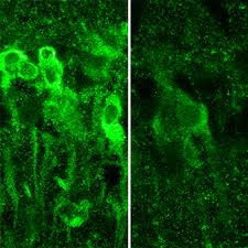
IMAGE:When exposed to large amounts of alcohol, neurons in the hippocampus produce steroids (shown in bright green, at left), which inhibit the formation of memory.
"It takes a lot of alcohol to block LTP and memory," says senior investigator Charles F. Zorumski, MD, the Samuel B. Guze Professor and head of the Department of Psychiatry. "But the mechanism isn't straightforward. The alcohol triggers these receptors to behave in seemingly contradictory ways, and that's what actually blocks the neural signals that create memories. It also may explain why individuals who get highly intoxicated don't remember what they did the night before."
But not all NMDA receptors are blocked by alcohol. Instead, their activity is cut roughly in half.
"The exposure to alcohol blocks some NMDA receptors and activates others, which then trigger the neuron to manufacture these steroids," Zorumski says.
The scientists point out that alcohol isn't causing blackouts by killing neurons. Instead, the steroids interfere with synaptic plasticity to impair LTP and memory formation.
"Alcohol isn't damaging the cells in any way that we can detect," Zorumski says. "As a matter of fact, even at the high levels we used here, we don't see any changes in how the brain cells communicate. You still process information. You're not anesthetized. You haven't passed out. But you're not forming new memories."
Stress on the hippocampal cells also can block memory formation. So can consumption of other drugs. When combined, alcohol and certain other drugs are much more likely to cause blackouts than either substance alone.
The researchers found that if they could block the manufacture of steroids by neurons, they also could preserve LTP in the rat hippocampus. And they did that with drugs called 5-alpha-reductase inhibitors. These include finasteride and dutasteride, which are commonly prescribed to reduce a man's enlarged prostate gland. In the brain, however, those substances seem to preserve memory.
"We would expect there may be some differences in the effects of alcohol on patients taking these drugs," Izumi says. "Perhaps men taking the drugs would be less likely to experience intoxication blackouts."
The researchers plan to study 5-alpha-reductase inhibitors to see how easily they get into the brain and to determine whether those drugs, or similar substances, might someday play a role in preserving memory.
Tokuda K, Izumi Y, Zorumski CF. Ethanol enhances neurosteroidogenesis in hippocampal pyramidal neurons by paradoxical NMDA receptor activation, The Journal of Neuroscience, vol. 31(27), pp. 9905-9909. July 6, 2011.
This work was supported by grants from the National Institute of Mental Health, the National Institute of General Medical Sciences, and the National Institute on Alcohol Abuse and Alcoholism of the National Institutes of Health (NIH), and by the Bantley Foundation.
Washington University School of Medicine's 2,100 employed and volunteer faculty physicians also are the medical staff of Barnes-Jewish and St. Louis Children's hospitals. The School of Medicine is one of the leading medical research, teaching and patient care institutions in the nation, currently ranked fourth in the nation by U.S. News & World Report. Through its affiliations with Barnes-Jewish and St. Louis Children's hospitals, the School of Medicine is linked to BJC HealthCare.
Labels:
induced,
memory,
neurons,
Social Worker Continuing Education
Subscribe to:
Posts (Atom)


